Caries
351 - Effect of Curodont Application on Oral Soft Tissue Toxicity

.jpg)
John C. Schmidt, Jr., DDS
PGY-2 Pediatric Dentistry Resident
Louisiana State University Health Sciences Center, New Orleans
New Orleans, Louisiana, United States- TL
Thomas Lallier, PhD
Professor, Cell Biology and Anatomy
LSUHSC School of Dentistry
New Orleans, Louisiana, United States - JJ
Jeffrey T. Johnson, DDS
Program Director
Louisiana State University
New Orleans, Louisiana, United States
Presenting Author(s)
Research Mentor(s)
Program Director(s)
Purpose: There are many options for treating early enamel or incipient carious lesions in the pediatric patient including fluoride varnish, SDF, and no treatment. Recently, a new product called Curodont has emerged as an alternative to more traditional approaches in managing incipient lesions. Previous research has indicated that Curodont is effective at remineralizing incipient lesions; however, there is no research on the product's effects on the surrounding soft tissue cells. The objective of this study was to evaluate the cytotoxicity of Curodont application on soft tissue concerning gingival fibroblasts by assessing survivability, proliferation, and attachment compared to SDF and fluoride varnish as controls. Knowing the potential cytotoxic effects of Curodont will allow clinicians to make educated decisions on its utilization in the treatment of early enamel or incipient carious lesions.
Methods: Assessing for proliferation, survivability, and attachment was accomplished by studying the effects of five varying concentrations of Curodont on gingival fibroblasts run in triplicate. Comparisons were made to optimal concentrations of SDF and fluoride varnish. Gingival fibroblasts were cell lines grown in vitro. Proliferation, survivability, and attachment were measured by a fluorescence microplate reader and an inverted fluorescence microscope.
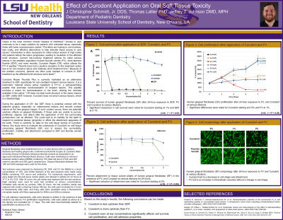
Results: There was a significant drop in cell survival for SDF at a concentration of 0.1%. Cell survival dropped to near 0% after 24 hours of exposure to SDF. SDF was not utilized in the cell adhesion or proliferation experiments because it caused cell death at such a low concentration. Curodont also had a significant drop in cell survival, starting at a concentration of 1%. A significant reduction in cell attachment was noted for Curodont starting at 2%. Cell proliferation was significantly affected for fluoride varnish at a concentration of 1% and for Curodont at 0.5% and 1%. Cell morphology visually explains the changes seen in the cell survival experiment. Curodont led to a decrease in cell shape and attachment, suggesting the cells were not surviving after treatment with Curodont. Fluoride varnish did not cause a change in cell shape, but an increase in intracellular vacuoles suggests that the cells are sickly but still alive.
Conclusions: Based on this study’s results, the following conclusions can be made: Curodont is less cytotoxic than SDF. Curodont is more toxic than FV. Curodont, even at low concentrations, significantly affects cell survival, cell proliferation, and cell adhesion properties.
Identify Supporting Agency and Grant Number:
Research supported by Louisiana State University School of Dentistry

.jpg)