Oral Pathology
320 - Juvenile Ossifying Fibroma: a Case Report with 5-year Follow-up

.jpg)
Andreia Infante, DDS, MSc
Postgraduate Student
Faculty of Dental Medicine of the University of Lisbon, Portugal- AJ
- IC
Ines Cardoso Martins, PhD
Faculty of Dental Medicine - University of Lisbon
- AC
Ana Coelho, PhD
Faculty of Dental Medicine - University of Lisbon
- PF
Paula Faria Marques, PhD
Head of Pediatric Dentistry Department
Faculty of Dental Medicine - University of Lisbon
LISBON, Lisboa, Portugal
Presenting Author(s)
Co-Author(s)
Program Director(s)
Introduction:
Juvenile ossifying fibroma (JOF) is a relatively rare benign fibro-osseous lesion that affects craniofacial skeleton of young patients, it’s a locally aggressive tumor with high recurrent potentials. May present as one of two histologic variants: Psammomatoid Juvenile Ossifying Fibroma (PsJOF) and Trabecular Juvenile Ossifying Fibroma (TrJOF). The TrJOF mainly affects the jaw and typically occurs in individuals aged 2-12. The growth of TrJOF is often described as painless, progressive, and sometimes rapid expansion of the affected area. Radiographically, these lesions could appear as a mixed radiolucent radiopaque areas usually expanding and well-defined, potentially resulting in cortical thinning or perforation.
Case Report:
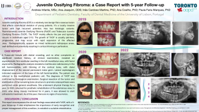
A 9-year-old female patient seeking for an evaluation due to dental crowding, without any other complaints and no significant medical history, was referred to our consultation. During the clinical examination, a firm vestibular swelling was noted in the left mandibular area, although the patient remained entirely asymptomatic. Additionally, facial asymmetry was observed. Radiographic analysis revealed a multilocular radiolucency in the left hemimandible, with thinning of the cortical bone. The radiograph also showed distal displacement of the germ of the second permanent molar. The lateral cephalogram indicated an expansion of the base of the left hemimandible. Subsequently, the patient was referred to the maxillofacial pediatric unit where appropriate diagnosis and treatment were initiated. This report encompasses the clinical findings associated with TrJOF, details the treatment and oral rehabilitation provided to our patient, and presents a 5-year follow-up. It also emphasizes the importance of early recognition and proper management.

.jpg)