Other
282 - Lead Shielding Effect on Scatter Radiation from Dental Radiographs


Lucy Knippenberg, DDS (she/her/hers)
Pediatric Dentistry Resident
Riley Hospital for Children
Riley Hospital for Children
Carmel, Indiana, United States- GE
George Eckert, MAS
Indiana University School of Dentistry
- BJ
Brandon Johnson, RDH, MS
Adams School of Dentistry Diagnostic Sciences
- JJ
James Jones, DMD, MSD, EdD, PhD
Riley Hospital for Children
- BS
Brian Sanders, DDS, MS
Riley Hospital for Children
- LV
LaQuia Vinson, DDS, MPH
Riley Hospital for Children
- PW
Phillip Wong, DMD
Indiana Univeristy School of Dentistry
- JY
Juan Yepes, DDS, MD, MPH, MS, DrPH, FDS, RCDS(Ed)
Riley Hospital for Children
.jpg)
Juan F. Yepes, DDS, MD, MPH, MS, DrPH, FDS, RCDS (ed)
Professor
Riley Hospital for Children
Indiana University -Riley Children Hospital-
Indianapolis, Indiana, United States- LV
LaQuia A. Vinson, DDS, MPH
Associate Professor
Indiana University, Bloomington, IN
Indianapolis, Indiana, United States
Presenting Author(s)
Co-Author(s)
Research Mentor(s)
Program Director(s)
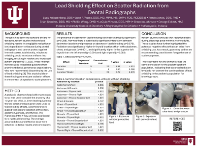
Purpose: Though it has been the standard of care for decades, recent studies indicate lead shielding results in a negligible reduction of ionizing radiation to tissues during dental radiographs and cannot protect against internal scatter. Additionally, misplaced shielding could introduce artifacts into imagery, resulting in retakes and increased patient exposure [1] [2] [3]. These findings have resulted in updated guidance from prominent dental governance organizations, who now recommend discontinuing the use of lead shielding [4]. This study builds on these findings to evaluate radiation effects in the context of a pediatric-scale phantom.
purpose of this study is to quantify the dose of scatter radiation at the level of the thyroid, chest, abdomen, and gonads with and without protective lead analogous shielding (thyroid collar/vest).
Methods: A pediatric phantom head with mannequin torso was used to model the anatomy of a 10-year-old-child. Dosimeters were placed at the level of the thyroid, chest, abdomen, and gonads. The Planmeca Intra X-Ray unit was positioned for a right side bitewing. The average absorbed dose and effective dose were obtained to derive results and conclusions.
Results: The presence or absence of lead shielding was not statistically significant (P = .288). Nor was there a statistically significant interaction between dosimeter location and presence or absence of lead shielding (P= .570).
Conclusions: Recent studies conclude that radiation doses during bitewings pose minimal risk to adults. These studies have further highlighted the potential negative effects that can arise from shielding use. As a result, governing bodies are recommending practitioners forego the use of such equipment.
This study tests for and demonstrates the same conclusion for the pediatric patient population, indicating that observed radiation levels do not warrant the continued use of lead shielding in the pediatric population for bitewing x-rays.
Identify Supporting Agency and Grant Number: Research supported by Indiana University Graduate Research Committee.

.jpg)