Caries
250 - Remineralization Effects of Salivary Vitamin D on Primary Dentition Caries


Noor M. Al Shaltoni, DDS
Resident
Boston University, Boston, MA
Boston, Massachusetts, United States- yF
yuwei Fan, PhD
Boston University
boston, Massachusetts, United States - KD
Keri Discepolo, DDS, MPH
Post Graduate Program Director of Pediatric Dentistry
Boston University Henry M. Goldman School of Dental Medicine
Boston, Massachusetts, United States
Presenting Author(s)
Research Mentor(s)
Program Director(s)
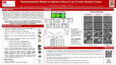
Methods: Thirty-one unidentified extracted human primary teeth were utilized for the purpose of this in-vitro study. Teeth were divided into 4 groups (n=8 teeth in groups A, C, D, n=7 teeth in group B) based on the concentration of vitamin D as follows; 1. artificial saliva without vitamin D as a control group, 2. artificial saliva with 20 nM of vitamin D to indicate deficiency, 3. artificial saliva with 40 nM of vitamin D to indicate insufficiency, and 4. artificial saliva with 100 nM of vitamin D to indicate sufficiency of vitamin D. Vickers microhardness testing (HV) was conducted before the initiation of artificial carious lesions to record the baseline. Subsequently, HV was recorded at 4 phases (n=7 teeth per group): at baseline, following immersion in artificial carious solution, midway through treatment cycling, and at the end of the 10-day experiment. Additionally, crystal morphology was examined using a field emission scanning electron microscope (FESEM) with different magnifications (n=1 tooth per group). Descriptive analysis and multiple factorial ANOVA used for data analysis. field emission scanning electron microscope (FESEM) with different magnifications (n=2 teeth per group). Results: The mean values of enamel surface microhardness exhibited a noticeable increase in groups C and D) compared to the group A and group B. Moreover, there is a statistically significant difference in enamel surface microhardness among groups B, C, D and control group A (p < 0.05). Additionally, examination under the FESEM revealed the presence of plate-like crystals on the enamel surface of teeth in groups A and C, whereas enamel surfaces treated with 100 nM VD3 (group D) displayed needle-like apatite crystallites.
Purpose: The aim of this in-vitro study is to investigate the remineralization effect of salivary vitamin D on artificially initiated carious lesions by analyzing changes in enamel microhardness and crystallization.
Conclusion: The application of Vitamin D demonstrated a notable enhancement in the densely crystallized enamel morphology and an improvement in surface microhardness. These findings emphasize the clinical significance of maintaining optimal concentrations of vitamin D in children for the effective remineralization of incipient carious lesions.
Identify Supporting Agency and Grant Number:

.jpg)