Oral Pathology
511 - Ameloblastic Fibro-Odontoma Case Report: An Interdisciplinary Team Approach


Kathryn M. Pawlak, DDS
Attending, University Pediatric Dentistry
University Pediatric Dentistry
University Pediatric Dentistry affiliated with University at Buffalo School of Dental Medicine
Buffalo, New York, United States- PC
Paul M. Creighton, DDS
University at Buffalo School of Dental Medicine

Kathryn M. Pawlak, DDS
Attending, University Pediatric Dentistry
University Pediatric Dentistry
University Pediatric Dentistry affiliated with University at Buffalo School of Dental Medicine
Buffalo, New York, United States
Presenting Author(s)
Co-Author(s)
Program Director(s)
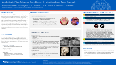
A 14-year-old male presented with chief complaint of a “lump” in the lower left mandible. The patient’s medical history was significant for ADHD.
Clinical examination revealed an extraoral expansion of the lower left mandible. Intraoral soft tissue swelling was not tender to palpation. Normal range of opening and no paresthesia was present.
Radiographic examination revealed a well-circumscribed and corticated lesion. The lesion presents as a mixed radiopaque/radiolucent multilocular lesion with honeycomb appearance. The lesion encompasses the body, ramus, coronoid and condylar process of the left mandible and extends to the apex of tooth 21. Tooth 18 is displaced mesially and inferiorly. Root blunting evident on tooth 20 and 21, however, no root resorption noted.
Patient was referred to OMFS where a biopsy was performed and sent for histological examination. This report includes the clinical and histological findings associated with Ameloblastic Fibro-Odontoma, surgical intervention and reconstruction rendered.
Identify Supporting Agency and Grant Number:

.jpg)